Notes
Let us observe the flowchart of co-ordination in a reflex system:
Stimulus -> Receptor -> Sensory neurone -> relay neurone in CNS -> motor neurone -> effector -> Response
Here we see that the brain has no function in the reflex system, thereby eliminating a stage in the response system in order to reduce time (i.e. no conscious decision).
Example of hot surface reaction:
Sensory organs contain receptors which, when stimulated by the intense heat, fire electrical impulses down through the spinal nerve. These impulses enter the CNS and are directly connected to the motor nerve by means of a relay nerve (this dramatically reduces response time and always produces the same response from the effector organs -> these are predetermined from DNA). This electrical impulse stimulates a response from the effector organ which in this case, would be to remove the sensory organ from the hot surface.
Note that reflexes are unconscious, always produce the same response, and are very rapid so as to reduce likelihood of organ or tissue damage.
Monday, 30 January 2012

2.84 understand that stimulation of receptors in the sense organs sends electrical impulses along nerves into and out of the central nervous system, resulting in rapid responses
Notes
Receptor organs (i.e. eyes) contain many neurones. These neurones, when acted upon by a stimuli, send electrical impulses across synapses and to other neurones which conduct this electrical information. This chain of neurones are known as nerves.
Once a receptor is stimulated, this electrical impulse is conducted down the spinal nerves to the spine. This is where it enters the CNS, from here, the electrical signal is interpreted by the brain, which sends another impulse to the effector organs, causing a response.
This all happens very rapidly due to the fast speed at which electrical energy can be transferred, which is the reason for our 'rapid responses'.
Receptor organs (i.e. eyes) contain many neurones. These neurones, when acted upon by a stimuli, send electrical impulses across synapses and to other neurones which conduct this electrical information. This chain of neurones are known as nerves.
Once a receptor is stimulated, this electrical impulse is conducted down the spinal nerves to the spine. This is where it enters the CNS, from here, the electrical signal is interpreted by the brain, which sends another impulse to the effector organs, causing a response.
This all happens very rapidly due to the fast speed at which electrical energy can be transferred, which is the reason for our 'rapid responses'.
2.83 recall that the central nervous system consists of the brain and spinal cord and is linked to sense organs by nerves
Notes
The CNS consists of:
a) the brain
b) the spinal cord
The sensory organs (receptors) send nerve impulses through the CNS to the effector organs (effectors) by means of nerves. A nerve consists of neurones (nerve cells, essentially) amongst other things.

The CNS consists of:
a) the brain
b) the spinal cord
The sensory organs (receptors) send nerve impulses through the CNS to the effector organs (effectors) by means of nerves. A nerve consists of neurones (nerve cells, essentially) amongst other things.

2.82 describe how responses can be controlled by nervous or by hormonal communication and understand the differences between the two systems
Notes
Nervous system refers to how the body responds to changes in the external (environment) and internal (organism) environment -> electrical impulses sent along the neurones to the CNS, which then decides activates the effectors and response.
The hormonal system refers to hormones as a means of communication between stimulus and response. Hormones are made in the endocrine glands which are then released directly into the blood and are carried by the circulatory system to the targeted effector muscles and organs.
The difference between the two is that since the endocrine gland is typically far away from the effector, it takes longer for the hormonal system to co-ordinate a response than the electrical impulse dependent nervous system. Also, since hormones stay in the circulatory system for a while after being secreted, the reactions they cause tend to be long-lasting.
Nervous system refers to how the body responds to changes in the external (environment) and internal (organism) environment -> electrical impulses sent along the neurones to the CNS, which then decides activates the effectors and response.
The hormonal system refers to hormones as a means of communication between stimulus and response. Hormones are made in the endocrine glands which are then released directly into the blood and are carried by the circulatory system to the targeted effector muscles and organs.
The difference between the two is that since the endocrine gland is typically far away from the effector, it takes longer for the hormonal system to co-ordinate a response than the electrical impulse dependent nervous system. Also, since hormones stay in the circulatory system for a while after being secreted, the reactions they cause tend to be long-lasting.
Monday, 16 January 2012
2.77a Thermoregulation
Notes
Homeostasis = "conditions are kept constant"
-> Homeothermic = "maintaining constant temperature (internal)"
-> e.g. Mammals are homeothermic organisms, carry out thermoregulation
Why do mammals want to preserve homeothermic conditions?
Due to enzyme reactions, attempt to keep temperature at 'optimum temperature' for most efficient rates of reactions - therefore body functions
-> Optimum temperature = homeothermic temperature of that organism
2.76 Sensitivity
Notes
Sensitivity = how organisms respond to changes in environment
Types of stimulus (change in environment):
-Light
-Temperature
-Pressure
-Chemicals
Detecting changes = receptors
Responding to changes = effectors (muscles/glands)
Saturday, 5 November 2011
2.75 Urine
Notes
Urine contains (excretion):
-> Salts (Affects composition of tissue fluid = osmoregulation)
-> Urea (Part of excretion of metabolic waste)
-> Water (Affects composition of tissue fluid = osmoregulation)
-> NO GLUCOSE
** Varies upon the conditions which a person is operating (environment/circumstances)


2.74 ADH
Notes
ADH = regulates water content of blood / control &alter composition of water in blood / more or less concentrated
--> Anti-diuretic hormone
--> Produced in hypothalamus
--> Flows through bloodstream --> kidney
**concentration must be right for osmoregulation (2.68b)
ADH targets collecting duct specifically
--> Collecting duct where water is selectively reabsorbed into bloodstream
--> ADH controls pores in the wall (more/less porous) to control 'flow' into bloodstream (more ADH ---> more porous)
Consequence of (more) ADH secretion
--> Urine more concentrated (more blood flows back into bloodstream)
--> Lower volume of urine
Questions?
ADH (hot day) = more ADH (more water available for sweat through the skin)
ADH (cold day) = less/no change ADH?
ADH (dehydrated) = more ADH (more water available for use in the body)


2.73 Glucose Re-absorption
Notes
Selective reabsorption of glucose occurs at PCT
--> Glomerula filtrate (glucose) ---reabsorption---> Blood stream
Filtrate contains:
-> Glucose
-> Water (collecting duct)
-> Salt
-> Urea
Urine = NO glucose? Due to filtration at PCT
--> Glucose is selectively reabsorbed (back into bloodstream) and taken out of the nephron system

2.72 Water Re-absorption
Notes
**Bowman's capsule = ultrafiltration ---> dissolved contents of plasma forced to become glomerula filtrate (water/salts/urea/glucose)
Filtration ---> filters out 'too much water' (as filtrate passes through nephron structure and reaches collecting duct)
---> Passing through collecting duct = water removed from filtrate (returned back to blood vessels/blood stream)
----> Water reselected to be reabsorbed into blood stream = 'selected reabsorption of water' occurs in collecting duct

2.71 Ultrafiltration
Notes
Nephron filters blood =
-> Filtered blood ('clean blood')
-> Excreted urine
----> Urine emerges from bottom of nephron (water / salts (NaCl,MgCl) / Urea (Toxic nitrogen waste)

How is urine formed? ULTRAFILTRATION (filtration of molecules)
1. Blood arrives into nephron through afferent arteriole (high blood pressure)
----> Blood vessel branches into coils (glomerulus)
----> Efferent arteriole (smaller diameter) = increase blood pressure flowing out (higher pressure)
**Glomerulus increases blood pressure
2. High pressure forces liquid in blood (plasma) into the inside of Bowman's capsule
----> Plasma contains:
-----------> water / salts / amino acids / glucose / urea)
----> Plasma inside capsule known as 'glomerula filtrate' (contains)

2.70 Nephron Structure
Notes
Process
Nephron = functional part of kidney (filtration/controlling composition of blood)
IN: Blood ---------- Renal artery (branch of aorta) --------> Nephron
OUT: Urine (excreted after filtration) <---- Ureter -------- Nephron
Urine collects in bladder for release
Filtered blood exits through renal vein ---> vena cava
Structure
-> Outer region = cortex {LIGHTER}
---> Inner region = medulla {DARKER}
--------> Inner space = pelvic region (where urine collects, drains down ureter)
**difference in colours = kidney is tubular structure made of tubes
Tube: starts on edge of medulla, moves outwards towards end of cortex, winds in cortex, dips down into medulla, back into cortex where winds again --> Dead end (Bowman's capsule)

Tubular structure (Nephron)
Twisted structure = convoluted tubules (tube)
Tube leading down to pelvic region (where urine is excreted) = Collecting ducts (tube)
Dip into medulla = loop of Henle
Dead end, cup shaped structure = Bowman's capsule
**tight knot of blood vessels (at Bowman's capsule) = glomerulus
* - - - u - - - /
1st convoluted tubule (- - -) = Proximal C.T. (PCT)
2nd convoluted tube (- - -) = Distal C.T (DCT)
---> Arrangement of nephron structure gives different colours of medulla & cortex
NOTE: millions of these structures (Nephrons) within a kidney.

Saturday, 29 October 2011
2.69 Urinary System - Describing the Structure
Notes
Urinary system
1. Kidneys (x2) (each with own blood supply / carries out excretion, osmoregulation
2. Ureter (x2) = tube from each kidney = carries urine from kidney to bladder
3. Bladder (x1) = stores urine
4. Urethra (x1) = urine to outside the body by tube through the (penis / vagina)

2.68b Osmoregulation
Notes
"Role of kidney in process of excretion an osmoregulation"
Osmo = 'osmosis'
Regulation = 'to control'
Cells in body
-> Tissue fluid (surrounding cells) = MUST be ISOTONIC with cytoplasm of cells
------> Water (In to cells) = Water (Out of cells) ---> Cells same size/shape, functions properly
**Danger to tissue (cells) by circulated blood**
-> Hypertonic = concentrated blood --> Remove too much water
-> Hypotonic = dilute blood --> Add too much water
ISOTONIC = Achieved by controlling composition of blood (forms tissue fluid) -> Role of kidneys
Blood circulates through kidney (in excess) are removed & excreted:
-> Salts = hypertonic
-> H2O = hypotonic
Excreted to control blood to be isotonic with cells cytoplasm -> maintains functions of cell & tissue

2.68a Excretion (Roles of the kidney & liver in excretion)
Notes
"Role of kidney in process of excretion and osmoregulation"
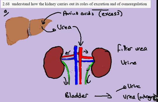
Excretion of Urea (contains nitrogen - toxic to body and cannot be stored)
-> Original form of nitrogen (circulatory system) = amino acids
-> EXCESS amino acids MUST be removed as TOXIC
-> Removal = role of liver & kidneys
PROCESS OF EXCRETION (of Urea)
1. Blood circulates to liver ---> amino acids broken down and converted --> Urea molecule
2. Urea circulates in blood stream -> Kidneys (both) --> Kidneys filter urea from blood
3. Urea + Water = Urine ----> Bladder (therefore removed from blood circulatory system
4. Filtered blood (from kidneys) returns to circulatory system
2.67b Human Organs of Excretion
Notes
1. Lungs
-> CO2
2. Kidneys
-> Salts
-> Urea (Nitrogen waste from amino acids -> amino acids not stored -> amino acids excreted)
-> Excess water
3. Skin
-> Salts (Sweat)
-> Urea (very small amounts)
-> Water (Sweat)

2.67a Excretion in Plants
Notes
1. DAY Leaf absorbs Light energy by Photosynthesis (metabolic reaction):
-> CO2 + H2O ------> C6H12O6 (glucose) + O2 (gas = waste molecule = excretion)
-> O2 metabolic waste = excreted
2. NIGHT Aerobic Respiration
-> C6H12O6 (glucose) + O2 ---Enzyme reactions (glucose broken down) ---> ATP + CO2 + H20
-> CO2 = metabolic waste = excreted
Excretion = Through stomatal pores

Friday, 14 October 2011
3.34 Causes of Mutation
Notes
Mutation
Gene (ACT) ---- mutation changes base sequence ----> New allele (AAT)
Causes
1. Ionising Radiation
-> X-rays / gamma rays
-> Ultraviolet: UV-B rays (sunshine) -> mutations such as skin cancer
2. Chemicals (mutagens = chemicals which cause mutations)
-> Tars (tobacco) -> cancerous conditions
* mutagens which also cause cancer = carcinogens

Mutation
Gene (ACT) ---- mutation changes base sequence ----> New allele (AAT)
Causes
1. Ionising Radiation
-> X-rays / gamma rays
-> Ultraviolet: UV-B rays (sunshine) -> mutations such as skin cancer
2. Chemicals (mutagens = chemicals which cause mutations)
-> Tars (tobacco) -> cancerous conditions
* mutagens which also cause cancer = carcinogens

3.33 Antibiotic resistance
Notes
Antibiotic resistance = increase in population of new allele
*see 3.31 evolution for details on MSSA/MRSA
MSSA --- random mutation (evolution) = MRSA---- process of natural selection ---> MRSA
As antibiotics used over time
-> MSSA: less common
-> MRSA: more common (resists antibiotic = population expands as normal) / antibiotic no longer 'works'

Antibiotic resistance = increase in population of new allele
*see 3.31 evolution for details on MSSA/MRSA
MSSA --- random mutation (evolution) = MRSA---- process of natural selection ---> MRSA
As antibiotics used over time
-> MSSA: less common
-> MRSA: more common (resists antibiotic = population expands as normal) / antibiotic no longer 'works'

3.32 Types of Mutation
Notes
Gene --- mutation ----> New Alleles
Alleles (responsible for phenotype) = harmful/beneficial/neutral
-> Beneficial = improve efficiency of enzyme
-> Harmful = production of non-functional enzyme
-> Neutral = second new allele / no effect AT PRESENT -> environmental change might render it beneficial or harmful
Gene --- mutation ----> New Alleles
Alleles (responsible for phenotype) = harmful/beneficial/neutral
-> Beneficial = improve efficiency of enzyme
-> Harmful = production of non-functional enzyme
-> Neutral = second new allele / no effect AT PRESENT -> environmental change might render it beneficial or harmful
3.31 Evolution
Notes
"Evolution by means of natural selection."
Evolution
- change in the form of organisms (new forms of organisms)
- change in frequency of alleles
Natural selection = mechanism of evolution (Charles Darwin)
Example:
Stapholococcus aureus = infection of skin, lungs (due to wounds e.g. operations)
-> Susceptible to being killed by Methecillin (Antibiotic)
S.A. which can be killed are known as the susceptible forms of the population
-> SUSCEPTIBLE: MSSA (methecillin susceptible s.a)
-> RESISTANT: Random Mutation to S.A. allows it to break down methecillin = no longer susceptible
When both forms are treated by methecillin (MSSA, MRSA)
-> SUSCEPTIBLE: less common, decrease in population
-> RESISTANT: more common, increase in population = increase in FREQUENCY of allele for resistance
*different forms = definition of evolution -> MRSA = evolved form of MSSA due to alterations in genome

PROCESS of NATURAL SELECTION
-> Random mutation = produces MRSA form
-> Non-random selection = applying antibiotics, MRSA selected for survival/MSSA
"Evolution by means of natural selection."
Evolution
- change in the form of organisms (new forms of organisms)
- change in frequency of alleles
Natural selection = mechanism of evolution (Charles Darwin)
Example:
Stapholococcus aureus = infection of skin, lungs (due to wounds e.g. operations)
-> Susceptible to being killed by Methecillin (Antibiotic)
S.A. which can be killed are known as the susceptible forms of the population
-> SUSCEPTIBLE: MSSA (methecillin susceptible s.a)
-> RESISTANT: Random Mutation to S.A. allows it to break down methecillin = no longer susceptible
When both forms are treated by methecillin (MSSA, MRSA)
-> SUSCEPTIBLE: less common, decrease in population
-> RESISTANT: more common, increase in population = increase in FREQUENCY of allele for resistance
*different forms = definition of evolution -> MRSA = evolved form of MSSA due to alterations in genome

PROCESS of NATURAL SELECTION
-> Random mutation = produces MRSA form
-> Non-random selection = applying antibiotics, MRSA selected for survival/MSSA
3.30 Mutation
Notes
Mutation = Rare, random change in genetic material that can be inherited
DNA -> Base sequence (gene) -> Form of gene = Allele
1. Changes in base sequence due to certain events
2. New version might result in production of NEW PROTEIN
3. Change in Phenotype
Dominant vs. Recessive = due to mutation, which changes base sequence of gene

Mutation = Rare, random change in genetic material that can be inherited
DNA -> Base sequence (gene) -> Form of gene = Allele
1. Changes in base sequence due to certain events
2. New version might result in production of NEW PROTEIN
3. Change in Phenotype
Dominant vs. Recessive = due to mutation, which changes base sequence of gene

Thursday, 13 October 2011
3.29 Species Variation
Notes
Variation = differences in phenotype of individual (possible to measure and show in graphic form)
Individual phenotype = Genotype + Modified by environment
Variation in Population = V P.genotype + V P.environment
-> Differences in Phenotypes of Species (appearance) = different genotypes + different habitats
Situations
1) Discontinuous Variation in Population/Species (GENOTYPE)
Variation in Genotype ONLY
-> Blood groups (O, A, A/B)

2) Continuous Variation in Population/Species (GENOTYPE + ENVIRONMENT)
Variation in Genotype + Environment
-> Height in humans = genes for height + diet

3) Variation in Population/Species (ENVIRONMENT)
Variation in Environment ONLY
-> Language spoken (not defined by genes at all) -> NOT INHERITABLE

Variation = differences in phenotype of individual (possible to measure and show in graphic form)
Individual phenotype = Genotype + Modified by environment
Variation in Population = V P.genotype + V P.environment
-> Differences in Phenotypes of Species (appearance) = different genotypes + different habitats
Situations
1) Discontinuous Variation in Population/Species (GENOTYPE)
Variation in Genotype ONLY
-> Blood groups (O, A, A/B)

2) Continuous Variation in Population/Species (GENOTYPE + ENVIRONMENT)
Variation in Genotype + Environment
-> Height in humans = genes for height + diet

3) Variation in Population/Species (ENVIRONMENT)
Variation in Environment ONLY
-> Language spoken (not defined by genes at all) -> NOT INHERITABLE

Sunday, 2 October 2011
3.18, 3.20, 3.21 Screen Capture
3.18c Co-dominance

3.21a Genetic Probabilities


3.21b Genetic Probabilities


3.20a Pedigree Diagrams

3.20b Pedigree Diagrams

Tuesday, 20 September 2011


Monday, 19 September 2011
3.2 Fertilisation
Notes
Fertilisation = "fusion of male and female gamete to produce a zygote that undergoes cell division and develops into an embryo"
1. In both Adult male & Adult female:
-> Each is in terms of a Diploid (2n) (diploid = complete set of chromosomes, 46)
-> Diploids divide in testis to produce gametes (meiosis = sperm and egg)
-> Diploid (2n) / 2 = Haploid (n)
-> 23 per sperm/egg
2. Fertilisation = two cells are fused together (Sperm gamete & egg gamete) to form ONE CELL
In human:
-> Haploid (n = 23) + Haploid (n = 23) => Diploid (2n = 46) = new cell known as ZYGOTE
-> Zygote combination of male and female cells but same no. of cells as adult human
3. Mitosis = cell divides (1-2-4-8-16, etc.), 2n -- mitosis --> 2n ALL CELLS CONTAIN DIPLOID (2n = 46) NUMBER OF CHROMOSOMES
-> Embryo = large collection of diploid cells
*Note: process is true for all sexually reproducing animals, variance in no. of chromosomes


3.9b Female Reproductive System
Notes
*before pregnancy, size of uterus = approx. orange
Ovary = meiosis (production of female gametes = eggs)
Oviducts (fallopian tube) = carries eggs to uterus BUT also location of where feritlisation may occur
*Fertilisation = occurs in oviducts NOT in uterus
Uterus = centre structure, wall of uterus which is a muscle stretches during pregnancy, contracts during birth
Lining of Uterus = develops fertilised eggs --> embryo --> child, placenta implants on wall of uterus
Uterus space = where embryo develops into unborn child, stretches to accommodate growing child
Cervix = where sperm cells from the penis enter, entrance to uterus, where sperm cells make way up to oviducts AND where eggs descend down towards
Vagina = collects sperm cells at top of vagina & allows sperm to enter through the cervix

3.9a Male Reproductive System
Notes
Bladder = stores urine
Testis = carry out meiosis produces gametes (sperm cells)
Epididymus = ('coil tube' system), stores sperm cells
Vas deferens = carry sperm cells to penis during sexual stimulation (during sex tube contracts and pushes sperm to penis)
Prostate = Adds 20% - 30% of volume of semen, contains sugars, alkali (alkali nature may neutralise acidic secretions vagina)
Seminal Vesicles = Adds remaining 70% of volume of semen so sperm can travel, produces sugar based secretion, alkali
*SEMEN = sperm cells + (seminal vesicle & prostate secretions)
Urethra = Runs length of penis, common tube joining R + L testis & vas deferens, exit for urine stored in bladder
Penis = Carry sperm cells into vagina during sexual intercourse

Saturday, 10 September 2011
3.12 Amniotic Fluid
Amniotic fluid: Protects developing embryo, surrounds embryo in uterus space
-> Fluid (protection of developing embryo)
-> Cannot be compressed,
-> Absorbs pressure when squeezed
= Prevents damage to embryo by absorbing forces on uterus wall
= Supports foetus as it cannot support own weight during development (bones = not calcified)
e.g. Trying to kick swiftly in water, but pressure (force) of kick is absorbed, slowing leg down

3.11 Placenta
Notes
Embryo: Unborn child, encompassed by uterus (amniotic fluid filled environment), with placenta on spinal side.
Placenta (provides nutrition to embryo)
-> Grows out of embryo in form of umbilical cord
-> Cord spreads out to form placental structure
-> Placenta grows into wall of uterus (past wall lining)
-> Glucose, amino acids & fats (maternal blood stream) --- crossing at (placenta) ---> embryo's blood stream
-> Efficiency/speed of blood/nutrition transfer:
-> Large S.A. of placenta
-> Very thin barrier (distance between) mother and embryo
-> Urea, carbon dioxide (embryo) --- crossing at (placenta) ---> maternal blood stream
** Placenta and placental blood vessels(including p. arteries & p. veins) belong biologically to embryo NOT mother


Subscribe to:
Posts (Atom)