Saturday 5 November 2011

2.75 Urine


Notes


Urine contains (excretion):

-> Salts (Affects composition of tissue fluid = osmoregulation)

-> Urea (Part of excretion of metabolic waste)

-> Water (Affects composition of tissue fluid = osmoregulation)

-> NO GLUCOSE


** Varies upon the conditions which a person is operating (environment/circumstances)


2.74 ADH


Notes


ADH = regulates water content of blood / control &alter composition of water in blood / more or less concentrated
--> Anti-diuretic hormone
--> Produced in hypothalamus
--> Flows through bloodstream --> kidney
**concentration must be right for osmoregulation (2.68b)

ADH targets collecting duct specifically
--> Collecting duct where water is selectively reabsorbed into bloodstream
--> ADH controls pores in the wall (more/less porous) to control 'flow' into bloodstream (more ADH ---> more porous)

Consequence of (more) ADH secretion
--> Urine more concentrated (more blood flows back into bloodstream)
--> Lower volume of urine

Questions?
ADH (hot day) = more ADH (more water available for sweat through the skin)
ADH (cold day) = less/no change ADH?
ADH (dehydrated) = more ADH (more water available for use in the body)


2.73 Glucose Re-absorption


Notes


Selective reabsorption of glucose occurs at PCT
--> Glomerula filtrate (glucose) ---reabsorption---> Blood stream

Filtrate contains:
-> Glucose
-> Water (collecting duct)
-> Salt
-> Urea

Urine = NO glucose? Due to filtration at PCT
--> Glucose is selectively reabsorbed (back into bloodstream) and taken out of the nephron system


2.72 Water Re-absorption


Notes


**Bowman's capsule = ultrafiltration ---> dissolved contents of plasma forced to become glomerula filtrate (water/salts/urea/glucose)

Filtration ---> filters out 'too much water' (as filtrate passes through nephron structure and reaches collecting duct)
---> Passing through collecting duct = water removed from filtrate (returned back to blood vessels/blood stream)
----> Water reselected to be reabsorbed into blood stream = 'selected reabsorption of water' occurs in collecting duct

2.71 Ultrafiltration


Notes


Nephron filters blood =
-> Filtered blood ('clean blood')
-> Excreted urine
----> Urine emerges from bottom of nephron (water / salts (NaCl,MgCl) / Urea (Toxic nitrogen waste)


How is urine formed? ULTRAFILTRATION (filtration of molecules)

1. Blood arrives into nephron through afferent arteriole (high blood pressure)
----> Blood vessel branches into coils (glomerulus)
----> Efferent arteriole (smaller diameter) = increase blood pressure flowing out (higher pressure)
**Glomerulus increases blood pressure

2. High pressure forces liquid in blood (plasma) into the inside of Bowman's capsule
----> Plasma contains:
-----------> water / salts / amino acids / glucose / urea)

----> Plasma inside capsule known as 'glomerula filtrate' (contains)

2.70 Nephron Structure


Notes


Process


Nephron = functional part of kidney (filtration/controlling composition of blood)

IN: Blood ---------- Renal artery (branch of aorta) --------> Nephron

OUT: Urine (excreted after filtration) <---- Ureter -------- Nephron

Urine collects in bladder for release
Filtered blood exits through renal vein ---> vena cava

Structure


-> Outer region = cortex {LIGHTER}
---> Inner region = medulla {DARKER}
--------> Inner space = pelvic region (where urine collects, drains down ureter)

**difference in colours = kidney is tubular structure made of tubes

Tube: starts on edge of medulla, moves outwards towards end of cortex, winds in cortex, dips down into medulla, back into cortex where winds again --> Dead end (Bowman's capsule)


Tubular structure (Nephron)

Twisted structure = convoluted tubules (tube)
Tube leading down to pelvic region (where urine is excreted) = Collecting ducts (tube)
Dip into medulla = loop of Henle
Dead end, cup shaped structure = Bowman's capsule
**tight knot of blood vessels (at Bowman's capsule) = glomerulus

* - - - u - - - /

1st convoluted tubule (- - -) = Proximal C.T. (PCT)
2nd convoluted tube (- - -) = Distal C.T (DCT)

---> Arrangement of nephron structure gives different colours of medulla & cortex

NOTE: millions of these structures (Nephrons) within a kidney.

Saturday 29 October 2011

2.69 Urinary System - Describing the Structure


Notes


Urinary system
1. Kidneys (x2) (each with own blood supply / carries out excretion, osmoregulation
2. Ureter (x2) = tube from each kidney = carries urine from kidney to bladder
3. Bladder (x1) = stores urine
4. Urethra (x1) = urine to outside the body by tube through the (penis / vagina)

2.68b Osmoregulation


Notes


"Role of kidney in process of excretion an osmoregulation"

Osmo = 'osmosis'
Regulation = 'to control'

Cells in body
-> Tissue fluid (surrounding cells) = MUST be ISOTONIC with cytoplasm of cells
------> Water (In to cells) = Water (Out of cells) ---> Cells same size/shape, functions properly

**Danger to tissue (cells) by circulated blood**
-> Hypertonic = concentrated blood --> Remove too much water
-> Hypotonic = dilute blood --> Add too much water

ISOTONIC = Achieved by controlling composition of blood (forms tissue fluid) -> Role of kidneys

Blood circulates through kidney (in excess) are removed & excreted:
-> Salts = hypertonic
-> H2O = hypotonic


Excreted to control blood to be isotonic with cells cytoplasm -> maintains functions of cell & tissue


2.68a Excretion (Roles of the kidney & liver in excretion)


Notes


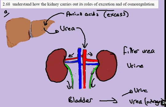
"Role of kidney in process of excretion and osmoregulation"

Excretion of Urea (contains nitrogen - toxic to body and cannot be stored)
-> Original form of nitrogen (circulatory system) = amino acids
-> EXCESS amino acids MUST be removed as TOXIC
-> Removal = role of liver & kidneys


PROCESS OF EXCRETION (of Urea)
1. Blood circulates to liver ---> amino acids broken down and converted --> Urea molecule
2. Urea circulates in blood stream -> Kidneys (both) --> Kidneys filter urea from blood
3. Urea + Water = Urine ----> Bladder (therefore removed from blood circulatory system
4. Filtered blood (from kidneys) returns to circulatory system

2.67b Human Organs of Excretion


Notes


1. Lungs 
-> CO2

2. Kidneys 
-> Salts
-> Urea (Nitrogen waste from amino acids -> amino acids not stored -> amino acids excreted)
-> Excess water

3. Skin
-> Salts (Sweat)
-> Urea (very small amounts)
-> Water (Sweat)

2.67a Excretion in Plants


Notes


1. DAY Leaf absorbs Light energy by Photosynthesis (metabolic reaction):
-> CO2 + H2O ------> C6H12O6 (glucose) + O2 (gas = waste molecule = excretion)
-> O2 metabolic waste = excreted

2. NIGHT Aerobic Respiration
-> C6H12O6 (glucose) + O2 ---Enzyme reactions (glucose broken down) ---> ATP + CO2 + H20
-> CO2 = metabolic waste = excreted

Excretion = Through stomatal pores

Friday 14 October 2011

3.34 Causes of Mutation

Notes


Mutation
Gene (ACT) ---- mutation changes base sequence ----> New allele (AAT)

Causes


1. Ionising Radiation 
   -> X-rays / gamma rays
   -> Ultraviolet: UV-B rays (sunshine) -> mutations such as skin cancer

2. Chemicals (mutagens = chemicals which cause mutations)
  -> Tars (tobacco) -> cancerous conditions

* mutagens which also cause cancer = carcinogens

3.33 Antibiotic resistance

Notes


Antibiotic resistance = increase in population of new allele

*see 3.31 evolution for details on MSSA/MRSA

MSSA --- random mutation (evolution) = MRSA---- process of natural selection ---> MRSA

As antibiotics used over time 
-> MSSA: less common

-> MRSA: more common (resists antibiotic = population expands as normal) / antibiotic no longer 'works'

3.32 Types of Mutation

Notes


Gene --- mutation ----> New Alleles

Alleles (responsible for phenotype) = harmful/beneficial/neutral

-> Beneficial = improve efficiency of enzyme

-> Harmful = production of non-functional enzyme

-> Neutral = second new allele / no effect AT PRESENT -> environmental change might render it beneficial or harmful

3.31 Evolution

Notes


"Evolution by means of natural selection."

Evolution
- change in the form of organisms (new forms of organisms)
- change in frequency of alleles

Natural selection = mechanism of evolution (Charles Darwin)

Example:

Stapholococcus aureus = infection of skin, lungs (due to wounds e.g. operations)
-> Susceptible to being killed by Methecillin (Antibiotic)

S.A. which can be killed are known as the susceptible forms of the population

-> SUSCEPTIBLE: MSSA (methecillin susceptible s.a)
-> RESISTANT: Random Mutation to S.A. allows it to break down methecillin = no longer susceptible

When both forms are treated by methecillin (MSSA, MRSA)

-> SUSCEPTIBLE: less common, decrease in population
-> RESISTANT: more common, increase in population = increase in FREQUENCY of allele for resistance

*different forms = definition of evolution -> MRSA = evolved form of MSSA due to alterations in genome


PROCESS of NATURAL SELECTION
-> Random mutation = produces MRSA form
-> Non-random selection = applying antibiotics, MRSA selected for survival/MSSA

3.30 Mutation

Notes


Mutation = Rare, random change in genetic material that can be inherited

DNA -> Base sequence (gene) -> Form of gene = Allele

1. Changes in base sequence due to certain events
2. New version might result in production of NEW PROTEIN
3. Change in Phenotype

Dominant vs. Recessive = due to mutation, which changes base sequence of gene

Thursday 13 October 2011

3.29 Species Variation

Notes


Variation = differences in phenotype of individual (possible to measure and show in graphic form)

Individual phenotype = Genotype + Modified by environment

Variation in Population = V P.genotype + V P.environment

-> Differences in Phenotypes of Species (appearance) = different genotypes + different habitats

Situations

1) Discontinuous Variation in Population/Species (GENOTYPE)

Variation in Genotype ONLY
-> Blood groups (O, A, A/B)


2) Continuous Variation in Population/Species (GENOTYPE + ENVIRONMENT)

Variation in Genotype + Environment
-> Height in humans = genes for height + diet


3) Variation in Population/Species (ENVIRONMENT)

Variation in Environment ONLY
-> Language spoken (not defined by genes at all) -> NOT INHERITABLE

Sunday 2 October 2011

3.18, 3.20, 3.21 Screen Capture

3.18c Co-dominance

3.21a Genetic Probabilities


3.21b Genetic Probabilities


3.20a Pedigree Diagrams

3.20b Pedigree Diagrams

Monday 19 September 2011

3.2 Fertilisation


Notes


Fertilisation = "fusion of male and female gamete to produce a zygote that undergoes cell division and develops into an embryo"

1. In both Adult male & Adult female:
-> Each is in terms of a Diploid (2n) (diploid = complete set of chromosomes, 46)
-> Diploids divide in testis to produce gametes (meiosis = sperm and egg)
    -> Diploid (2n) / 2 = Haploid (n)
    -> 23 per sperm/egg

2. Fertilisation = two cells are fused together (Sperm gamete & egg gamete) to form ONE CELL

In human:
-> Haploid (n = 23) + Haploid (n = 23) => Diploid (2n = 46) =  new cell known as ZYGOTE
-> Zygote combination of male and female cells but same no. of cells as adult human

3. Mitosis = cell divides (1-2-4-8-16, etc.), 2n -- mitosis --> 2n ALL CELLS CONTAIN DIPLOID (2n = 46) NUMBER OF CHROMOSOMES
-> Embryo = large collection of diploid cells

*Note: process is true for all sexually reproducing animals, variance in no. of chromosomes


3.9b Female Reproductive System


Notes

*before pregnancy, size of uterus = approx. orange

Ovary = meiosis (production of female gametes = eggs)

Oviducts (fallopian tube) = carries eggs to uterus BUT also location of where feritlisation may occur

*Fertilisation = occurs in oviducts NOT in uterus

Uterus = centre structure, wall of uterus which is a muscle stretches during pregnancy, contracts during birth

Lining of Uterus = develops fertilised eggs --> embryo --> child, placenta implants on wall of uterus

Uterus space = where embryo develops into unborn child, stretches to accommodate growing child

Cervix = where sperm cells from the penis enter, entrance to uterus, where sperm cells make way up to oviducts AND where eggs descend down towards

Vagina = collects sperm cells at top of vagina & allows sperm to enter through the cervix

3.9a Male Reproductive System


Notes


Bladder = stores urine

Testis = carry out meiosis produces gametes (sperm cells)

Epididymus = ('coil tube' system), stores sperm cells

Vas deferens = carry sperm cells to penis during sexual stimulation (during sex tube contracts and pushes sperm to penis)

Prostate = Adds 20% - 30% of volume of semen, contains sugars, alkali (alkali nature may neutralise acidic secretions vagina)

Seminal Vesicles = Adds remaining 70% of volume of semen so sperm can travel, produces sugar based secretion, alkali

*SEMEN = sperm cells + (seminal vesicle & prostate secretions)

Urethra = Runs length of penis, common tube joining R + L testis & vas deferens, exit for urine stored in bladder

Penis = Carry sperm cells into vagina during sexual intercourse

Saturday 10 September 2011

3.12 Amniotic Fluid


Notes


Amniotic fluid: Protects developing embryo, surrounds embryo in uterus space

-> Fluid (protection of developing embryo)
     -> Cannot be compressed, 
     -> Absorbs pressure when squeezed
= Prevents damage to embryo by absorbing forces on uterus wall
= Supports foetus as it cannot support own weight during development (bones = not calcified)

e.g. Trying to kick swiftly in water, but pressure (force) of kick is absorbed, slowing leg down

3.11 Placenta


Notes


Embryo: Unborn child, encompassed by uterus (amniotic fluid filled environment), with placenta on spinal side.

Placenta (provides nutrition to embryo)


-> Grows out of embryo in form of umbilical cord

-> Cord spreads out to form placental structure

-> Placenta grows into wall of uterus (past wall lining)

-> Glucose, amino acids & fats (maternal blood stream) --- crossing at (placenta) ---> embryo's blood stream

-> Efficiency/speed of blood/nutrition transfer:
   -> Large S.A. of placenta
   -> Very thin barrier (distance between) mother and embryo

-> Urea, carbon dioxide (embryo) --- crossing at (placenta) ---> maternal blood stream


** Placenta and placental blood vessels(including p. arteries & p. veins) belong biologically to embryo NOT mother